Evaluating Kidney Stone Treatment Options: How Specialist Care Protects Renal Function
Introduction
A sudden ache in the lower back or side often serves as the first warning sign that mineral deposits have built up in the urinary tract. While minor urinary crystals frequently pass without major disruption, larger deposits can block fluid flow, trigger sharp discomfort, and raise the risk of urinary tract infections. Recognizing early indicators helps individuals decide when home monitoring is sufficient and when specialist intervention is necessary.Resolving renal stones involves a range of strategies, from dietary modifications and oral medications to targeted procedures like shock wave therapy or endoscopic laser surgery. Learning how urologists pinpoint stone location and evaluate renal health makes deciding on a care plan much simpler. Resources like
Understanding Kidney Stones and Urological Health
Renal stones, clinically known as nephrolithiasis or urolithiasis, develop when dissolved minerals in urine aggregate into solid masses inside the kidneys. The urinary system—comprising the kidneys, ureters, bladder, and urethra—relies on adequate fluid volume to flush out metabolic waste. When concentration levels rise, substances like calcium oxalate, uric acid, or phosphate bind together into crystalline structures.
These deposits vary in size, from sub-millimeter grains that exit unnoticed to multi-centimeter structures that plug the narrow ureteral passage. Anyone can form stones, but hydration levels, metabolic activity, and anatomical variations heavily influence individual vulnerability. Seeking timely diagnostic checks prevents complications such as severe urinary blockages, localized tissue inflammation, or secondary bacterial growth.
Common Symptoms and Warning Signs
Small stone fragments occasionally travel through the urinary tract without causing noticeable discomfort. However, once a deposit shifts position or restricts urine flow within the ureter, physical symptoms become distinct:
Sharp, intermittent flank pain: Discomfort typically originates in the lower back or side, often radiating toward the groin and lower abdomen.
Urinary urgency and dysuria: A persistent desire to urinate, accompanied by burning or discomfort during voiding.
Hematuria (blood in urine): Urine that displays pink, reddish, or brownish hues caused by internal tissue friction.
Turbid or cloudy urine: Off-colored or strong-smelling urine, which can signal co-existing bacterial activity.
Gastrointestinal distress: Nausea or episodes of vomiting caused by shared nerve pathways between the kidneys and abdominal organs.
Because symptoms like burning voiding or pelvic pressure overlap with conditions such as a urinary tract infection (UTI) or an enlarged prostate (BPH), a professional clinical evaluation is required for an accurate diagnosis.
Causes and Risk Factors
Mineral crystallization in the renal system rarely stems from a single trigger. Instead, multiple physiological and environmental variables contribute to stone formation over time:
Chronic fluid deficits: Insufficient daily liquid intake leaves urine concentrated, accelerating crystal aggregation.
Dietary imbalances: Consuming high levels of sodium, animal protein, or oxalate-heavy foods elevates mineral excretion into the urine.
Genetic predisposition: A personal history or family background of renal stones raises long-term recurrence rates.
Metabolic and digestive disorders: Conditions such as hyperparathyroidism, chronic diarrhea, or malabsorption syndromes alter normal calcium and uric acid processing.
Pharmacological factors: Long-term use of specific medications, including certain antacids, diuretics, or high-dose supplements, can disrupt urine chemistry.
Diagnosis and Diagnostic Tests
Urology specialists utilize targeted physical assessments, lab work, and medical imaging to evaluate stone size, composition, and location before creating a care plan.
Clinical history intake: Reviewing symptom duration, dietary patterns, and underlying medical conditions.
Urinalysis: Testing urine samples for microscopic blood cells, elevated mineral levels, pH balance, and signs of bacterial infection.
Metabolic blood panels: Checking serum calcium, uric acid, and creatinine levels to assess overall renal function.
Non-contrast computed tomography (CT): High-resolution CT scans provide 3D imagery to confirm exact stone dimensions and location. Ultrasound serves as a valuable, non-radiative alternative for pregnant patients or routine monitoring.
Crystalline analysis: Laboratory evaluation of retrieved stone fragments to determine chemical makeup and guide long-term prevention strategies.
Testing strategies are customized based on symptom severity, patient medical history, and clinical presentation.
Kidney Stone Treatment Options
Choosing a treatment path depends on stone dimensions, location within the urinary tract, pain intensity, and whether fluid drainage from the kidney is impaired.
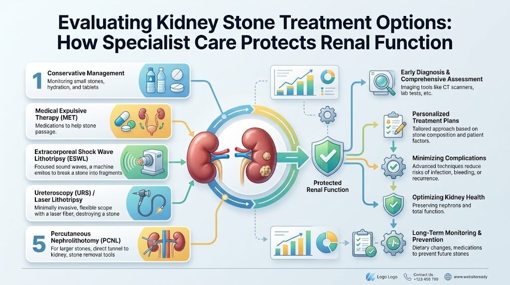
Conservative Management and Observation
For small deposits (generally under 5 millimeters) that do not obstruct urine flow or trigger severe symptoms, conservative strategies are often sufficient:
Fluid therapy: Consuming high volumes of water to encourage natural passage through the ureter.
Symptom management: Utilizing prescribed or over-the-counter anti-inflammatory medications to control localized pain.
Medical Expulsive Therapy (MET): Using selective alpha-blockers to relax smooth muscle tissue in the ureter, allowing stone fragments to pass faster with less resistance.
Non-Invasive Procedures
Extracorporeal Shock Wave Lithotripsy (ESWL): Directs focused acoustic waves from outside the body to break stones in the kidney or upper ureter into tiny, passables particles.
Minimally Invasive Procedures
Ureteroscopy (URS): Involves inserting a thin, flexible scope equipped with optics through the urethra and bladder into the ureter. A holmium or thulium laser breaks the stone down, and tiny extraction baskets retrieve the fragments.
Percutaneous Nephrolithotomy (PCNL): Reserved for complex or large stones (typically over 2 centimeters). The specialist creates a small incision in the patient's back to access the kidney directly, using nephroscopes and ultrasonic or pneumatic tools to fragment and remove the stone.
Robotic Urology Surgery and Advanced Reconstructive Care
In rare cases involving significant anatomical abnormalities, severe ureteral strictures, or massive staghorn stones, open or robotic-assisted reconstructive procedures may be necessary to restore normal urinary drainage and remove dense mineral deposits.
Urology Hospitals and Urology Specialists
Selecting an appropriate clinical facility plays a vital role in managing complex stone cases and other urological concerns. Specialized urology centers offer advanced imaging, modern endoscopic equipment, and experienced care teams.
When selecting a healthcare provider, evaluate these key factors:
Specialist qualifications: Ensure board-certified urologists have extensive experience in endoscopic, shock wave, and percutaneous stone techniques.
Facility capabilities: Confirm the hospital offers full diagnostic imaging, laser fragmentation tools, and minimally invasive surgical suites.
Urgent care access: Verify the hospital can manage emergency obstructions, acute renal colic, or stone-related infections promptly.
Preventive follow-up care: Access to metabolic evaluations, dietary counseling, and routine monitoring helps minimize recurrence risks.
For patients seeking local care options, searching for a "urologist near me" or exploring platforms like
Comparison of Kidney Stone Interventions
| Treatment Option | Primary Application | Potential Advantages | Key Considerations |
| Observation & Hydration | Small stones (<5mm), minimal pain | Completely non-invasive, low cost | Requires time; patient must manage temporary discomfort |
| Medical Expulsive Therapy | Moderate stones, no infection | Relaxes ureteral walls to ease passage | Requires prescription; potential mild side effects like dizziness |
| Shock Wave Lithotripsy (ESWL) | Uncomplicated kidney/upper ureter stones | Outpatient procedure, no incision required | May require repeat sessions if stones do not fully fragment |
| Ureteroscopy (URS) | Mid-to-lower ureteral and kidney stones | High success rate, direct visual control | Performed under anesthesia; may require temporary stent placement |
| Percutaneous Nephrolithotomy (PCNL) | Large (>2cm) or staghorn stones | Clears dense, large stone burdens efficiently | Involves a tiny back incision, brief hospital stay, and recovery period |
Practical Patient Examples
Example 1: Managing Small Stones Conservatively
A 38-year-old individual experiences mild side pain. A low-dose CT scan reveals a 4mm stone in the lower ureter without urinary blockage or infection. The attending urologist recommends a conservative plan: increasing daily water intake, taking anti-inflammatory pain relievers, and using an alpha-blocker to relax the ureteral smooth muscle. The stone passes naturally five days later without invasive treatment.
Example 2: Addressing Recurrent Obstructive Pain
A 50-year-old individual presents with acute flank pain, vomiting, and trace blood in their urine sample. Diagnostic imaging reveals a 9mm stone lodged in the upper ureter, causing fluid buildup in the left kidney. Given the stone size and ongoing blockage, the urologist performs a laser ureteroscopy under general anesthesia, safely fragmenting the stone and restoring normal urinary flow.
Decision-Making Framework for Patients
[ Step 1: Identify Physical Symptoms ]
          │
          ▼
[ Step 2: Diagnostic Evaluation ] ──► (CT Imaging / Ultrasound / Urinalysis)
          │
          ▼
[ Step 3: Assess Stone Profile ] ──► (Check Size, Location, and Density)
          │
          ▼
[ Step 4: Consult Urology Specialist ] ──► (Compare Conservative vs. Surgical Interventions)
          │
          ▼
[ Step 5: Select Facility & Provider ]
          │
          ▼
[ Step 6: Execute Treatment Plan & Preventive Metabolic Monitoring ]
Common Mistakes to Avoid
Ignoring ongoing side or back pain: Delaying evaluation for persistent abdominal or flank discomfort can lead to severe kidney infections or long-term renal damage.
Self-diagnosing online: Assuming severe side pain is just a routine muscle strain or minor stone without proper clinical imaging or urine testing.
Using unprescribed antibiotics: Taking leftover antibiotics for urinary discomfort without confirming whether an infection or stone obstruction is present.
Assuming surgery is mandatory: Overlooking conservative monitoring or non-invasive options for small, uncomplicated stones.
Skipping metabolic follow-up: Failing to analyze passed stone fragments or complete blood work, missing key insights to prevent future stone formation.
Practical Tips / Key Takeaways
Maintain consistent fluid intake: Drink enough fluids throughout the day to keep urine pale and diluted, minimizing mineral crystallization.
Seek early medical advice: Schedule an evaluation promptly if you experience severe back pain, discolored urine, or burning during voiding.
Explore all treatment pathways: Discuss conservative, non-invasive, and surgical options with your physician to select the right approach for your stone profile.
Catch and save passed stone fragments: Use a urine strainer to collect stone pieces for laboratory analysis, helping your specialist design a targeted prevention plan.
Complete scheduled follow-up visits: Attend post-treatment imaging sessions to confirm full stone clearance and ensure healthy kidney function.
Frequently Asked Questions (FAQs)
1. What does a urologist treat?
A urologist is a medical specialist focused on conditions affecting the male and female urinary systems, as well as the male reproductive organs. Their scope of care includes managing kidney stone treatment, addressing enlarged prostate concerns, treating complex infections, and overseeing specialized therapies for prostate cancer treatment or bladder cancer treatment.
2. When should someone see a urologist for kidney stones?
Consult a urologist if you experience severe flank pain, discolored urine, persistent nausea, or trouble voiding. Emergency medical attention is required if symptoms include high fever, chills, or a complete inability to pass urine.
3. How are kidney stones diagnosed?
Physicians diagnose renal stones using physical examinations, urine analysis, blood testing, and medical imaging. Non-contrast CT scans or ultrasounds help pinpoint the exact size, position, and structural impact of the stone within the urinary tract.
4. Can kidney stones pass on their own?
Yes, smaller stones (typically under 5 millimeters) often pass naturally through the urinary tract. Drinking plenty of water and taking prescribed muscle-relaxing medications can help ease passage.
5. What non-invasive kidney stone treatments are available?
Extracorporeal Shock Wave Lithotripsy (ESWL) is a common non-invasive option. It uses targeted shock waves generated outside the body to break stones into small fragments, which can then exit through urine naturally.
6. How does ureteroscopy work for kidney stone treatment?
Ureteroscopy involves guiding a small scope through the urethra and bladder directly into the ureter or kidney. The specialist uses a laser to break the stone apart and extracts the pieces using tiny retrieval instruments, avoiding external incisions.
7. What is the difference between BPH and prostate cancer?
Benign Prostatic Hyperplasia (BPH) is a non-cancerous enlargement of the prostate gland common in older men. While BPH and prostate cancer share similar lower urinary tract symptoms, they are distinct conditions evaluated using PSA testing, exams, and imaging.
8. When is robotic urology surgery considered?
Robotic urology surgery is typically reserved for complex anatomical reconstructions, intricate partial nephrectomies, or advanced oncological procedures. It offers enhanced 3D visual precision, though candidacy depends on the patient's health status and condition complexity.
9. How can I find accredited urology hospitals?
Look for facilities with board-certified specialists, modern imaging, advanced surgical equipment, and structured emergency protocols. Patient platforms like UrologyHospitals.com simplify this search by providing detailed information on provider capabilities and treatment centers.
10. How do I choose a urologist near me?
When searching for a local specialist, review their clinical experience, subspecialization, hospital affiliations, diagnostic tools, and care options. Evaluating credentials helps ensure you select a provider suited to your specific health needs.
Conclusion
Managing renal stones begins with recognizing early warning signs, obtaining accurate diagnostic imaging, and selecting an appropriate care plan. Whether a stone requires routine monitoring, oral medication, shock wave lithotripsy, or endoscopic laser surgery, working alongside an experienced urology specialist ensures safe and effective management.Taking proactive steps protects overall renal function and reduces the risk of recurrent stone formation. Resources like UrologyHospitals.com help patients understand conditions, research modern treatment options, and connect with reputable healthcare providers—enabling informed decisions throughout every stage of care.